
A new organelles have been discovered in human cells, and scientists call them “semi-hardened.”
Like our body’s full-size organs, the intracellular organelles are specialized structures that perform certain functions. Observing the filaments that preserve the shape of the cells, University of Virginia Assistant Professor Seham Ebrahim and her team noticed a new structure that is consistently appearing in the 3D images they are creating.
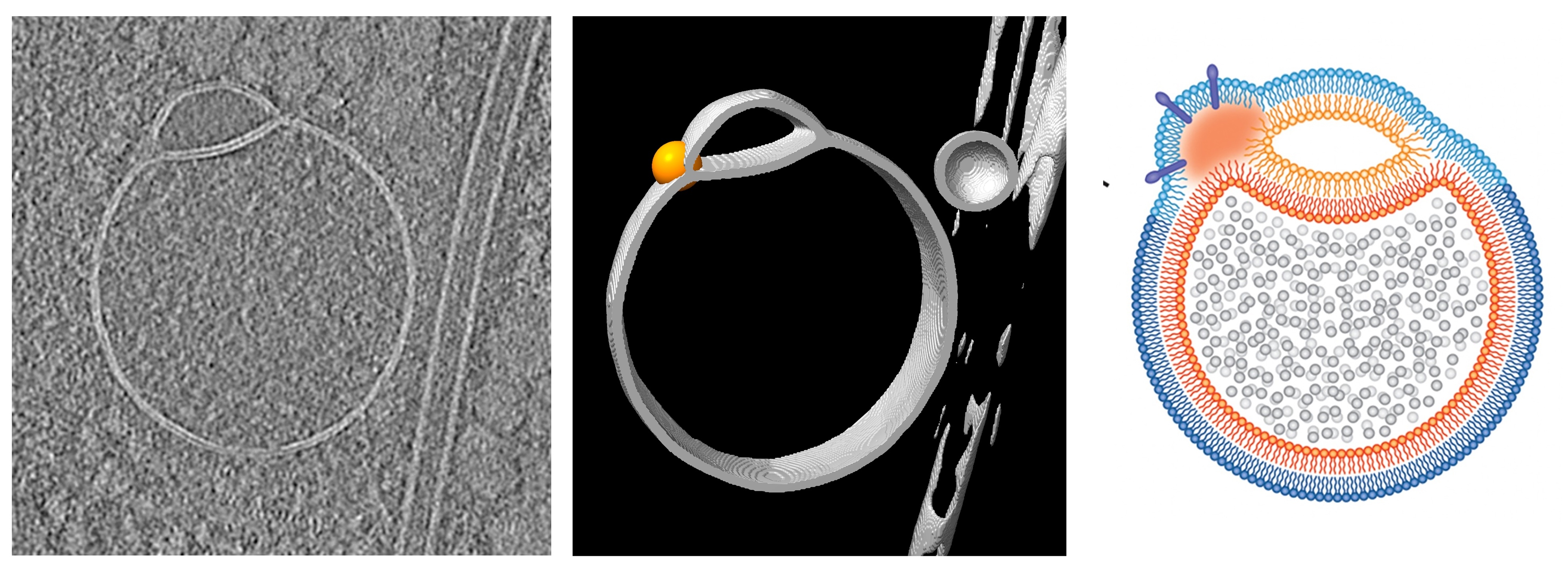
At first, what appeared to be an artifact in the image was found to be a new organelles that could be involved in sorting, recycling and disposing of proteins in human cells. Ebrahim compared the hemifsome shape to the shape of a scarfed snowman. Imagine a small head with a small head attached to a large body. A thin boundary separates both ends.
You might like it
The organelles have a diameter of about 100 nanometers, less than half of the small mitochondria, the cell’s famous powerhouse.
Scientists were able to observe semispores because they used a method called cryo-electron tomography (cryo-et) to generate images. This technique rapidly freed cells from four lab-grown cell lines, allowing them to preserve as many structures as possible and create clear 3D images.
“It’s like a snapshot of time without chemicals or dirt types,” Ebrahim told Live Science. Using this imaging technique, she said they can see inside the cells in “very native state” as if they were glass balls.
Related: Meet the new organelles, “Frodosome”
In their paper published in the journal Nature Communications in May, the researchers wrote a harsh processing procedure that other imaging techniques are likely to prevent cluster cells from being observed previously. Furthermore, in other techniques using imaging to study traffic deployed in live cells, organelles were probably too small to see and appeared as a blur at best, Ebrahim added.
Ebrahim and her colleagues were seeing the composition of vesicles they had never observed before. Vesicles are balloon-like structures that are used to transport proteins, hormones, and more intracellularly and between cells. A new study revealed two vesicles and two vesicles fused with two layers of fat barrier.
“From a biophysics perspective, it’s a breakthrough,” Ebrahim said. This observation influenced the name semidual maturation, as one side refers to a partial merger of two bilayers.
Ebrahim claims that semispores can be classified as organelle because they are self-contained functional units within cells, in contrast to the “flanked” structures that appear temporarily as membranes formed and divided. In her paper, she wrote that it was an unlikely cryo-et artifact.
Ebrahim’s findings suggest that the half they visualize is not a freezing-induced distortion, but a real cell intermediate,” says Yi-Wei Chang, assistant professor of biochemistry and biophysics at the University of Pennsylvania Perelman University School of Medicine, who is not involved in the work.
Once the role and function of blood semiuniformity is confirmed through further research, they could be recognized as a unique class of intermediate structures that meet some of the mammalian cell fusion process, Chan told Live Science in an email.
Current research allows researchers to confirm that semi-hardening is present, but have not yet determined the exact role, life cycle, or composition of organelles. Ebrahim assumes that the semispore membrane is a precursor for a particular type of vesicle. She believes that semispores can play an important role in recycling or disposing of cell membranes.
Researchers write that it could unleash new insights into how the hemisphere works and how diseases such as Alzheimer’s disease are becoming sick. Alzheimer’s disease is associated with inappropriate clearance of abnormal protein plaques in the brain and accumulates over time.
“Without cryoelectron tomography, we would have missed this discovery,” Ebrahim said.
Source link