simple facts
Milestone: Dissection of the famous patient “Tan”
Date: April 18, 1861
Location: Bicêtre Hospital on the outskirts of Paris
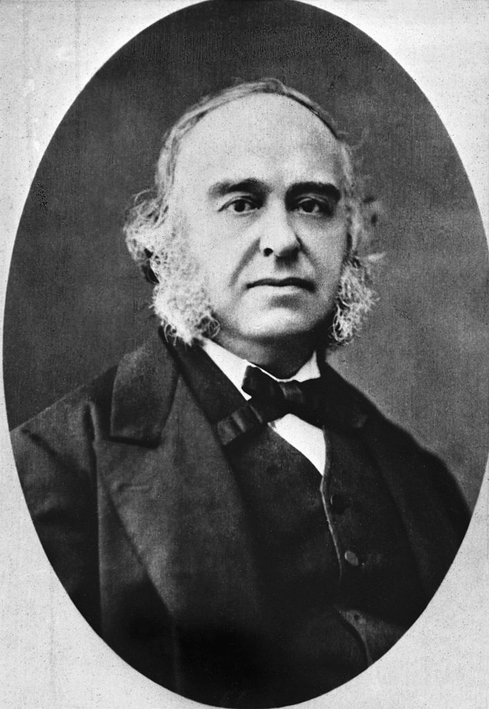
People: Dr. Paul Broca
On April 18, 1861, a doctor in Paris cut open the brain of a patient who had died the previous day and unwittingly identified the brain regions key to spoken language.
The patient, Louis-Victor Leborgne, was nicknamed “Tan” by the doctors at the Bicêtre hospital because it was one of the only words he could say. Before his death at the age of 51, he spent 21 years in a hospital’s psychiatric ward.
Leborgne was reportedly healthy at birth, but began having epileptic seizures as an infant. At the age of 30, he lost the ability to speak. For some time he avoided medical treatment, but was eventually admitted to Bicêtre Hospital.
Article continues below
you may like
Doctors discovered that he understood language well and could use gestures to communicate his needs. On rare occasions, he would swear.
Ten years after admission, he began to experience paralysis on his right side, which gradually worsened, and he also experienced mental difficulties. Eventually he lost the ability to walk. He spent the last seven years of his life in bed.
Over the past few years, Dr. Paul Broca, a surgeon at the hospital, has come to see Leborgne as a patient.
According to the translation, Mr. Broca said of his patient: “His most common numeric reaction was by opening and closing his fingers. He could tell the time on the clock to the second without error. He knew exactly how many years he had been in Bicêtre, etc.”
“However, many questions that a person of ordinary intelligence would have found a way to answer with gestures remained unanswered in an intelligible manner. In other cases, the responses were clear but did not answer the question,” Broca observed. “Undoubtedly, the patient’s intelligence was affected to a considerable extent, but he certainly maintained more intelligence than was necessary for conversation.”
On April 17, 1861, Leborgne died of gangrene. It was probably caused by pressure sores on my feet. The next day, Broca began the dissection and noticed a pocket of clear fluid about the size of a “hen’s egg” in the perisylvian region of the left hemisphere of the brain. This region surrounds a deep groove called the lateral sulcus, which marks the upper border of the temporal lobe. Some areas around the fluid showed “softness”. There were other abnormalities as well. Leborgne’s brain was lighter than normal, and some areas of the brain had smaller volumes than expected.
On the same day, Broca presented the results of the autopsy at the Anthropological Society Congress in Paris. At the time, there was an ongoing debate between scientists who believed that all functions of the brain were distributed throughout the organ system, and those who believed that specific areas performed specific functions.
What to read next
Broca’s autopsy provided strong evidence for the latter idea.
“The main site and natural location of tenderness is the central part of the frontal lobe of the left hemisphere. That’s where we find the most extensive lesions, the most advanced and oldest lesions,” he said in his presentation.
This “suggests that frontal lobe damage is responsible for the language loss in this case,” Broca added.
However, at the conference, colleagues did not immediately recognize the significance of this discovery. Much of the conference focused on the now-discredited racial “science” that links skull measurements to intelligence. However, by August 1861, Broca had studied the brains of several patients with what would later be called aphasia. This research strengthened his belief that speech was localized in the frontal lobe, which he later narrowed to the left frontal lobe.
Throughout his career, Broca not only identified the areas involved in aphasia, but also noted that speech therapy sometimes helped patients recover their language.
Since Broca’s time, researchers have identified distinct brain regions that perform specific cognitive functions and have focused on more precise areas of the brain key to speech than Broca identified. This area, now named Broca’s area, is recognized as important in Broca’s aphasia, where patients can understand language but have difficulty producing spoken, written, or signed language.
We now know that other regions and networks beyond the realm of brokers play a major role in language. For example, damage to Wernicke’s area, discovered in 1874, can cause a type of aphasia in which patients speak in long, complete sentences that have little meaning.
Leborgne’s intact brain, which Broca examined only on the surface without cutting it for decades, was on view at the Dupuytren Museum in Paris, which closed to the public in 2016.
Source link